
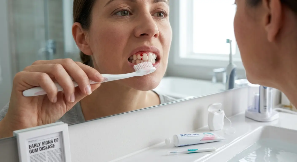
Gum disease begins with signs that most patients have learned to ignore: a little pink in the sink when brushing, gums that look slightly puffy, persistent bad breath that mints don’t fix. None of these things hurt, and that is exactly why they are dangerous, the disease is silent at the stage where it is still completely reversible, and it is loud at the stage where it is no longer reversible at all.
This guide explains how gum disease actually progresses, what the early signs mean, when the disease crosses from reversible (gingivitis) to non-reversible (periodontitis), and what the modern, evidence-based treatment pathway involves at each stage.
The short version: gum disease has two main stages. Gingivitis (the early stage) is fully reversible with professional cleaning and good home care. Periodontitis (the advanced stage) involves loss of the bone that holds the teeth in place, it can be controlled, but the lost attachment doesn’t come back on its own. The earlier you catch it, the more you keep.
What gum disease actually is
“Gum disease” is a non-clinical umbrella term for two related but distinct conditions: gingivitis and periodontitis. They are not the same disease at different volumes, they are biologically and clinically different, and the line between them matters.
Gingivitis: inflammation without attachment loss
Gingivitis is inflammation of the gum tissue caused by bacterial plaque accumulation along the gumline. The clinical signs are redness, swelling, and bleeding on brushing or probing. The defining feature is what is not there: no loss of the connective tissue attachment between the tooth and the bone, and no loss of the supporting alveolar bone underneath.
Because nothing has been lost yet, gingivitis is fully reversible. Removal of the bacterial trigger, through professional cleaning combined with consistent home plaque control, returns the tissue to a healthy baseline within roughly two weeks. The current global classification recognizes plaque-induced gingivitis as a distinct diagnostic category, with several modifying factors (hormonal changes, certain medications, smoking) that can amplify the response without changing the fundamental reversibility [1].
Periodontitis: when the supporting structures begin to fail
Periodontitis is what happens when the inflammation of gingivitis is sustained long enough to spread into the deeper structures that hold the tooth in place, the periodontal ligament and the alveolar bone. The bacterial composition of the biofilm shifts toward anaerobic species adapted to the low-oxygen environment under the gumline, including the well-characterized red complex described by Socransky and colleagues: Porphyromonas gingivalis, Tannerella forsythia, and Treponema denticola [2].
In response, the host immune system mounts an inflammatory cascade involving pro-inflammatory cytokines (IL-1β, TNF-α, IL-6), prostaglandins, and matrix metalloproteinases (MMP-8, MMP-9). These molecules degrade collagen and stimulate osteoclast activity, leading to the loss of periodontal attachment and the resorption of supporting bone that defines the disease. The bone that is lost in periodontitis does not regenerate spontaneously after the inflammation is controlled, which is why the line between gingivitis and periodontitis is the most clinically important line in periodontal medicine.
In simpler terms, gingivitis is inflammation that hasn’t damaged anything structural yet, and it can be undone. Periodontitis is the same inflammation that has reached the bone, and it leaves a permanent mark on the support around the tooth.
How common is gum disease? The numbers most patients haven’t heard
Gum disease is far more prevalent than most patients assume. The CDC, using data from the National Health and Nutrition Examination Survey (NHANES), has consistently reported that approximately 42 percent of U.S. adults aged 30 years and older have some form of periodontitis, with prevalence rising to over 70 percent in adults aged 65 and older [3]. Severe periodontitis affects roughly 7 to 9 percent of the adult population. Gingivitis, the precursor stage, is even more common, it has been documented in over half of the adult population at any given assessment.
Periodontitis is the leading cause of tooth loss in adults in the United States. It is also one of the most preventable. Most cases that progress to advanced disease pass through years of recognizable warning signs first.
The early signs you can recognize at home
The signs below are the ones a patient is most likely to notice without a clinical examination. None of them resolves without intervention, and most progress predictably if ignored. Recognizing any of them is the appropriate moment for evaluation, not when one of the late signs (mobility, drift, pus, deep recession) appears.
Bleeding when you brush, floss, or probe with the tongue
Healthy gum tissue does not bleed during routine plaque control. Any bleeding is a clinical finding that almost always reflects gingivitis (early, reversible) or periodontitis (with attachment loss). Patients often interpret bleeding as a sign that they are brushing or flossing “too hard” and reduce the intensity, the opposite of what is needed. The bleeding stops when the bacterial trigger is removed, not when the mechanical cleaning is reduced.
Simply explained, if your gums bleed when you brush or floss, treat it as a clinical signal, not a normal variation. The intervention required at the bleeding stage is far less than what becomes necessary if the same disease progresses.
Red, puffy, or shiny gums where they should look firm and pink
Healthy gingival tissue has a coral-pink color in most patients (with normal variation in patients with darker pigmentation), a firm consistency, and a stippled texture similar to orange peel along the attached gingiva. Inflamed gums lose the stippling, take on a more uniform shiny appearance, develop a deeper red or purplish hue, and feel softer to the touch. The change is often gradual and easy to miss in the mirror unless you actively look for it.
Color and texture changes alone are enough to warrant a clinical evaluation. They reflect the same underlying inflammatory cascade as bleeding, even when bleeding has not yet become noticeable.
Gums that look longer, or teeth that look longer than they used to
Gingival recession is the apical migration of the gum margin, exposing more of the tooth root. The clinical signs the patient sees are teeth that look longer, dark triangles between teeth where the gum used to fill the space, increased sensitivity at the gumline, and root surfaces that catch food. Recession can occur in patients with healthy gums (from aggressive brushing, traumatic occlusion, or thin tissue biotype) or as a consequence of periodontal attachment loss, and the distinction matters because the underlying causes are different.
In practical terms, any new recession, particularly when accompanied by bleeding or sensitivity, deserves clinical evaluation. Recession does not reverse on its own and tends to progress without intervention.
Persistent bad breath that brushing doesn’t fix
Chronic halitosis that persists through good oral hygiene is rarely gastrointestinal in origin. The dominant chemistry is the production of volatile sulfur compounds, hydrogen sulfide, methyl mercaptan, dimethyl sulfide, by anaerobic bacteria in periodontal pockets and on the tongue. The same bacterial species that drive periodontitis are heavy producers of these compounds, which is why halitosis correlates with periodontal status.
Persistent bad breath is a clinical finding with clinical causes, not a cosmetic concern. A periodontal evaluation and a thorough professional cleaning resolve most cases. Mouthrinses mask the symptom temporarily but do not address the source.
Loose teeth, drifting teeth, or a bite that has changed
These signs are no longer early. By the time a patient notices that a tooth feels loose, that the front teeth have drifted apart, or that the bite has shifted, periodontal attachment loss is typically substantial. Any new mobility, drift, or change in occlusion deserves prompt evaluation. The likelihood that the affected teeth can be saved is strongly time-dependent at this stage.
Pus or persistent localized swelling around a tooth
Localized swelling at the gumline, with or without spontaneous drainage of pus, suggests an active periodontal abscess. The clinical scenario is typically a deep periodontal pocket that has become acutely infected, sometimes painful, sometimes painless if drainage relieves pressure. Periodontal abscesses respond predictably to drainage and local debridement, but they signal that the underlying disease is advanced and requires comprehensive periodontal management.
How gum disease is staged and graded today
The 2018 World Workshop on the Classification of Periodontal and Peri-Implant Diseases produced the current global classification system used in modern dental practice. It replaces the older system of “mild, moderate, severe” with a more clinically useful framework based on stage and grade [4].
Staging: how much support has been lost
The stage describes the severity and complexity of the disease at the time of diagnosis. It is based on the amount of clinical attachment loss measured at the worst-affected site, the radiographic bone loss, and the number of teeth lost from periodontitis. The four stages:
- Stage I (initial periodontitis): 1-2 mm of clinical attachment loss, less than 15 percent radiographic bone loss, no tooth loss from periodontitis.
- Stage II (moderate periodontitis): 3-4 mm of clinical attachment loss, 15-33 percent radiographic bone loss, no tooth loss from periodontitis.
- Stage III (severe periodontitis with potential for additional tooth loss): 5 mm or more of clinical attachment loss, more than 33 percent bone loss, up to 4 teeth lost from periodontitis, and complications such as deep pockets, vertical bone defects, or furcation involvement.
- Stage IV (advanced periodontitis with extensive tooth loss and potential masticatory dysfunction): the same severity findings as stage III plus 5 or more teeth lost from periodontitis and complications affecting bite function.
Grading: how fast the disease is progressing
The grade describes the rate of progression and incorporates risk factors that predict future loss. Three grades:
- Grade A (slow rate): no evidence of bone or attachment loss over 5 years; non-smoker; no diabetes.
- Grade B (moderate rate): less than 2 mm of bone loss over 5 years; smoker fewer than 10 cigarettes per day; HbA1c less than 7.0% in patients with diabetes.
- Grade C (rapid rate): more than 2 mm of bone loss over 5 years; smoker 10 or more cigarettes per day; HbA1c 7.0% or higher in patients with diabetes.
In practical terms: stage and grade together describe what has happened and how fast it is happening. A patient with stage II grade C disease has lost a moderate amount of bone but is losing it rapidly, and that combination predicts a different treatment intensity than stage II grade A. The classification is not academic; it determines the treatment plan and the recall interval.
When gum disease is reversible, and when it isn’t
The single most important question for any patient who has been told they have gum disease is whether the changes can be reversed. The honest answer depends entirely on the stage.
Gingivitis is fully reversible
Plaque-induced gingivitis is fully reversible by the removal of the bacterial trigger. After a professional cleaning to disrupt and remove established biofilm and calculus, and consistent twice-daily plaque control at home, gingival inflammation resolves within approximately two weeks in most patients. The tissue color, contour, and bleeding tendency return to baseline. Nothing has been lost, and nothing needs to be rebuilt.
This is the entire reason why catching gum disease at the gingivitis stage matters so much. The intervention is the simplest thing dentistry offers, a cleaning and behavioral conversation, and the outcome is a return to full health. Once the disease has progressed past gingivitis, that simplicity disappears.
Periodontitis can be controlled, but lost attachment doesn’t come back on its own
Periodontitis is, by definition, characterized by loss of clinical attachment and supporting bone. With evidence-based treatment, inflammation can be controlled, the bacterial load reduced, and the disease stabilized indefinitely. With consistent maintenance after treatment, patients with periodontitis can keep their teeth for the rest of their lives at rates approaching those of patients who never had periodontitis. What treatment cannot do is regenerate the bone and connective tissue that were lost during the active disease, except in specific defect morphologies amenable to regenerative procedures.
The clinical implication: the goal of periodontal therapy in established disease is to stop further loss, not to recover what is gone. The earlier that treatment begins, the less there is to lose, which translates directly into better long-term tooth retention and less need for restorative or implant care later.
Treatment by stage: what each step actually involves
The 2020 European Federation of Periodontology S3 clinical practice guideline on the treatment of stage I-III periodontitis is the current evidence-based framework used internationally and at D&FD. It defines a stepwise approach in which each step builds on the success of the previous one [5].
Step 1: behavior change and supragingival management
The first step of periodontal therapy applies to all patients regardless of stage and addresses the foundation: oral hygiene instruction, removal of plaque retentive factors (overhanging restorations, ill-fitting prostheses, deep carious lesions), professional supragingival biofilm and calculus removal, and risk factor management (tobacco cessation, glycemic control, dietary guidance). The success of this step is reassessed before moving forward, because patient adherence is the single strongest predictor of long-term outcomes.
Step 2: subgingival instrumentation (the core of periodontal therapy)
Step 2 addresses the subgingival environment where periodontitis is sustained. Subgingival instrumentation, historically called scaling and root planing, is performed under local anesthesia and removes calculus and disrupted biofilm from the root surfaces within the periodontal pocket. The procedure is performed quadrant by quadrant or more efficiently full-mouth, depending on the case, using ultrasonic instrumentation, hand instrumentation with periodontal curettes, or a combination.
In selected cases, adjunctive therapies are added at this step, systemic antibiotics for specific clinical phenotypes (typically generalized stage III grade C periodontitis), locally delivered antimicrobials in deep residual pockets, or sub-antimicrobial-dose doxycycline as a host modulator. The choice is individualized; routine antibiotic use is not part of standard periodontal therapy.
Reassessment occurs approximately 6 to 8 weeks after subgingival instrumentation. Sites that respond, reduced probing depths, no bleeding on probing, transition to maintenance. Sites that remain non-responsive (residual pockets above 5 mm with bleeding) are considered for the next step.
Step 3: surgical and regenerative therapy for non-responsive sites
Step 3 applies only to specific sites that did not resolve with non-surgical therapy. The treatment options include access flap surgery (open-flap debridement to gain visual access to deep root surfaces), resective surgery to recontour bone defects, and regenerative procedures designed to rebuild lost periodontal tissue in defect morphologies that allow it. Regenerative options include guided tissue regeneration with barrier membranes, enamel matrix derivative (Emdogain), and bone replacement grafts, each indicated in specific defect anatomies.
Surgical periodontal therapy is technique-sensitive and outcome-dependent on case selection. The 2020 EFP guideline emphasizes that the goal is to manage residual disease in a small, defined subset of sites, not to surgically treat every periodontitis case [5].
Step 4: supportive periodontal care for life
Step 4 is the maintenance protocol that follows active treatment and continues indefinitely. It includes professional supportive periodontal care at three- or four-month intervals (the EFP S3 guideline recommends three months as the default), full periodontal re-evaluation at every visit, reinforcement of home care, and re-treatment of any sites that show signs of recurrence.
The evidence for maintenance is one of the most consistent findings in periodontology. Patients on regular supportive periodontal care after treatment retain their teeth at rates close to those of patients who never had periodontitis. Patients who skip maintenance or revert to a routine six-month prophylaxis schedule show progressive disease recurrence and tooth loss [5,6]. Maintenance is not optional after periodontal therapy; it is the treatment continuing at a reduced frequency.
Why gum disease matters beyond your mouth
Periodontitis is increasingly recognized as a chronic systemic inflammatory load with measurable effects on health outside the mouth. The associations are not folklore, they are quantified in large prospective cohort studies and meta-analyses, and several have been characterized at the mechanistic level.
Cardiovascular disease: a documented association
A 2023 meta-analysis of 39 prospective cohort studies including 4.4 million individuals reported that periodontitis was associated with a 24 percent increased risk of major adverse cardiovascular events (RR 1.24, 95% CI 1.15-1.34), a 20 percent increased risk of coronary heart disease (RR 1.20, 95% CI 1.12-1.29), a 26 percent increased risk of stroke (RR 1.26, 95% CI 1.15-1.37), and a 42 percent increased risk of cardiac death (RR 1.42, 95% CI 1.10-1.84) [7]. The proposed mechanism is shared chronic systemic inflammation: bacterial products and inflammatory mediators from periodontal pockets contribute to the systemic inflammatory milieu that drives endothelial dysfunction and atherosclerotic plaque progression. Whether the relationship is causal or shared-pathway remains an active area of research, but the association is robust.
Treatment of periodontitis has been shown to improve several markers of cardiovascular risk, most notably C-reactive protein, an inflammatory marker tracked clinically in cardiovascular medicine, within months of non-surgical periodontal therapy [8]. The clinical inference is that controlling periodontal inflammation may have benefits beyond the mouth, particularly in patients already at elevated cardiovascular risk.
Diabetes: a bidirectional relationship
Periodontitis and type 2 diabetes influence each other through documented inflammatory and metabolic mechanisms covered in detail in our companion satellite on cleaning frequency. The short version: hyperglycemia amplifies the inflammatory response to subgingival biofilm, and untreated periodontal inflammation worsens glycemic control. Non-surgical periodontal therapy lowers HbA1c by an average of 0.3 to 0.4 percentage points at three to four months, a clinically meaningful effect comparable to adding a second oral hypoglycemic agent in some patients [9,10].
Basically, patients with diabetes need shorter periodontal recall intervals (three to four months), and the periodontal exam is one of the few diagnostic tools that can flag a need for re-evaluation of glycemic control.
Pregnancy outcomes, respiratory disease, and rheumatoid arthritis
Periodontitis has been associated with adverse pregnancy outcomes (preterm birth, low birth weight) in multiple observational studies, although intervention trials have produced mixed results regarding whether treatment changes those outcomes. Maintaining periodontal health during pregnancy remains the consensus recommendation, both for the reasons specific to pregnancy-related gingivitis and for the broader inflammatory load argument.
Associations have also been documented between periodontitis and aspiration pneumonia in older or hospitalized patients, between periodontitis and rheumatoid arthritis (with shared mechanisms involving citrullination), and between periodontitis and several other chronic inflammatory conditions. The strength of these associations varies, and causality remains difficult to establish, but the consistent direction across diseases reinforces the principle that controlling oral inflammation has health implications beyond dentistry.
Preventing recurrence after treatment
The risk of recurrence after periodontal treatment is real, and it is the reason maintenance is structured as a permanent protocol rather than a course of treatment. Several factors have been consistently identified as risk factors for recurrence in the periodontology literature.
Risk factors for periodontal disease recurrence
- Inadequate home care: the strongest single predictor of recurrence. Plaque control, not professional intervention, determines what happens between maintenance visits.
- Continued tobacco use or vaping: smokers have measurably worse response to periodontal therapy and higher recurrence rates than non-smokers. Vaping has now been documented to carry similar periodontal risk.
- Poorly controlled diabetes: HbA1c above target predicts both faster initial progression and higher recurrence rates after treatment.
- Failure to convert from prophylaxis to periodontal maintenance: returning to a routine six-month cleaning after periodontal therapy is one of the most consistent predictors of recurrence in the long-term cohort literature.
- Untreated parafunctional habits: bruxism and clenching that exceed the periodontium’s capacity to adapt accelerate attachment loss in already weakened sites.
- Medication-related factors: medications that cause hyposalivation, gingival overgrowth (calcium channel blockers, cyclosporine, phenytoin), or impaired healing can elevate recurrence risk.
In practical terms, the patient who finishes active periodontal therapy and stops there has not finished the disease management. The patient who finishes therapy, attends three-month maintenance, controls plaque well, addresses systemic risk factors, and reports new symptoms early has the realistic prospect of keeping the affected teeth indefinitely.
How D&FD approaches gum disease at every stage
Periodontal evaluation is part of every comprehensive examination at D&FD, performed by our doctors. The data set generated at every visit, full-mouth probing depths at six sites per tooth, bleeding on probing, recession measurements, mobility grading, furcation involvement, and keratinized tissue width, establishes the baseline at the first visit and identifies trends over time at every subsequent one.
The clinical philosophy at D&FD is preservation-first: the earliest, least invasive intervention that can stop disease is always preferred over a more involved intervention at a later stage. For gum disease, that means recognizing gingivitis at the visit it appears, and treating it before it becomes periodontitis. For patients who already have periodontitis, it means staging and grading the disease accurately, treating it according to the EFP S3 guideline framework, and committing to the maintenance protocol that keeps it controlled.
The same principle connects this article to the broader implant cluster. Many patients who eventually need bone grafting before implant placement lost the original tooth to untreated periodontitis. The bone that will need to be rebuilt with a graft is often bone that could have been preserved with periodontal therapy years earlier.
If you have noticed any of the early signs in this guide, bleeding gums, persistent bad breath, gum recession, color or texture changes, a comprehensive periodontal evaluation is the appropriate next step. Both D&FD offices, Gables Dental Care (3815 SW 8th Street, Coral Gables) and EliteDent (12538 SW 120th Street, Kendall), follow the same protocols and any of our dentists can see you at either location.
References
[1] Chapple ILC, Mealey BL, Van Dyke TE, et al. Periodontal health and gingival diseases and conditions on an intact and a reduced periodontium: Consensus report of workgroup 1 of the 2017 World Workshop on the Classification of Periodontal and Peri-Implant Diseases and Conditions. J Periodontol. 2018;89(Suppl 1):S74–S84.
[2] Socransky SS, Haffajee AD, Cugini MA, Smith C, Kent RL Jr. Microbial complexes in subgingival plaque. J Clin Periodontol. 1998;25(2):134–144.
[3] Eke PI, Thornton-Evans GO, Wei L, Borgnakke WS, Dye BA, Genco RJ. Periodontitis in US adults: National Health and Nutrition Examination Survey 2009-2014. J Am Dent Assoc. 2018;149(7):576–588.
[4] Tonetti MS, Greenwell H, Kornman KS. Staging and grading of periodontitis: Framework and proposal of a new classification and case definition. J Periodontol. 2018;89(Suppl 1):S159–S172.
[5] Sanz M, Herrera D, Kebschull M, et al. Treatment of stage I-III periodontitis: The EFP S3 level clinical practice guideline. J Clin Periodontol. 2020;47(Suppl 22):4–60.
[6] Trombelli L, Franceschetti G, Farina R. Effect of professional mechanical plaque removal performed on a long-term, routine basis in the secondary prevention of periodontitis: A systematic review. J Clin Periodontol. 2015;42(Suppl 16):S221–S236.
[7] Guo X, Li X, Liao C, Feng X, He T. Periodontal disease and subsequent risk of cardiovascular outcome and all-cause mortality: A meta-analysis of prospective studies. PLOS ONE. 2023;18(9):e0290545.
[8] Meng R, Xu J, Fan C, et al. Effect of non-surgical periodontal therapy on risk markers of cardiovascular disease: A systematic review and meta-analysis. BMC Oral Health. 2024;24(1):692.
[9] Sanz M, Ceriello A, Buysschaert M, et al. Scientific evidence on the links between periodontal diseases and diabetes: Consensus report and guidelines of the joint workshop on periodontal diseases and diabetes by the IDF and the EFP. J Clin Periodontol. 2018;45(2):138–149.
[10] Simpson TC, Clarkson JE, Worthington HV, et al. Treatment of periodontitis for glycaemic control in people with diabetes mellitus. Cochrane Database Syst Rev. 2022;(4):CD004714.