
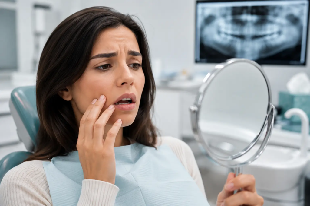
The signs that most reliably predict a serious dental problem rarely involve pain. By the time a tooth hurts, the disease underneath it is usually advanced, and the treatment that would have been a small filling six months earlier has often become a root canal, a crown, or an extraction. The early signals show up in the everyday details: bleeding when you brush, new sensitivity to cold, gums that look longer than they used to, a tooth that feels slightly different when you bite down, persistent bad breath, a change in how a tooth looks, jaw clicking, or a mouth that feels chronically dry.
This guide is a complete clinical reference to the warning signs of dental disease, what each one means, what causes it, what the dentist looks for, and when the situation has crossed from “book a visit soon” to “call now.”
The short version, most dental diseases are silent in their early stages. Pain is a late signal. Early detection through scheduled examinations and recognition of the asymptomatic warning signs in this guide is what separates a small intervention from a major one, in clinical and financial terms.
Why pain is a late signal in dentistry
Three of the most common dental diseases progress without symptoms until they are clinically advanced. Understanding why is the foundation for understanding the rest of this guide.
The neuroanatomy of dental pain
Dental enamel is not innervated. Dentin contains odontoblastic processes that respond to fluid movement within the dentinal tubules, the mechanism behind the cold sensitivity associated with hydrodynamic theory, but the response is dampened in early lesions and only becomes prominent when the lesion approaches the pulp. The pulp itself is well innervated, but the nerve fibers (mostly A-delta and unmyelinated C fibers) tend to produce significant symptoms only when the inflammation has progressed to the stage of acute pulpitis or apical pathology. By that point, the lesion is usually no longer minimally invasive to treat.
Periodontal disease is even more clinically silent. The destruction of connective tissue attachment and alveolar bone in periodontitis is mediated by host-derived inflammatory pathways (cytokines such as IL-1β, TNF-α, and IL-6, and matrix metalloproteinases such as MMP-8 and MMP-9) responding to subgingival bacterial pathogens, the so-called red complex of Porphyromonas gingivalis, Tannerella forsythia, and Treponema denticola [1]. The process produces no nociceptive signal. The first symptoms a patient typically perceives are mobility, drift, or change in occlusion, all late-stage findings.
Oral and oropharyngeal cancers compound the same problem. Most early lesions are painless and easily missed without a structured screening. Five-year relative survival is approximately 88 percent when the cancer is detected at a localized stage and falls to roughly 41 percent at distant stage [2]. The clinical implication is that detection at the asymptomatic stage materially changes prognosis.
The economic logic of early detection
A carious lesion detected at the demineralization stage is reversible with topical fluoride and home-care modification. The same lesion at the dentin stage requires a small composite filling. At the dentino-pulpal complex it requires endodontic therapy and indirect restoration. Once the tooth is non-restorable it requires extraction, replacement, and, if bone has resorbed, grafting before implant placement. The cost difference between the first and last steps in this progression routinely exceeds the order of magnitude. The same logic applies across periodontal, restorative, and surgical care.
Early warning signs and what each one actually means
The signs below all warrant clinical evaluation. None is an emergency on its own, but none resolves without intervention, and most progress predictably if ignored.
Bleeding when you brush or floss
Healthy gingival tissue does not bleed during routine mechanical plaque control. Bleeding on brushing or flossing almost always indicates one of two diagnoses: gingivitis, defined by inflammation without loss of clinical attachment and reversible with proper professional and home care, or periodontitis, defined by inflammation with documented attachment loss and non-reversible without active therapy.
The clinical distinction is made by periodontal probing. Probing depths of 3 mm or less without bleeding represent health. Probing depths of 4 mm or more, or any depth with bleeding on probing, indicate active disease. Generalized bleeding on probing in more than 20 percent of sites is a marker of generalized inflammation and elevated risk of progression [3]. Gingivitis typically resolves within two weeks of professional cleaning and consistent home care; periodontitis requires staged non-surgical periodontal therapy [3].
In practical terms, bleeding gums are not normal, and they are not something to wait out. The intervention required at the bleeding stage is far less than what becomes necessary when the same disease progresses to mobility or attachment loss.
New or persistent sensitivity to cold, heat, or sweet stimuli
Dentinal sensitivity has multiple potential etiologies, and the differential matters because the treatment depends on the cause. The most common causes in adult patients are: gingival recession exposing root dentin, enamel erosion from chronic acid exposure, incipient interproximal caries, occlusal wear and abfraction lesions secondary to bruxism, microcracks in the enamel from parafunctional loading, and reversible pulpitis from a deep restoration.
Sensitivity that is brief and tied to a specific stimulus (a sip of cold water, a sweet bite) is usually peripheral. Sensitivity that lingers after the stimulus is removed (more than 10 to 15 seconds), wakes the patient at night, or radiates may indicate irreversible pulpitis and requires urgent assessment. Sensitivity localized to one specific tooth, especially with a pulsatile or throbbing quality, suggests pulpal involvement rather than peripheral dentinal exposure.
This means that a single new sensitivity that resolves quickly may be peripheral and benign, but persistent, lingering, or worsening sensitivity is a clinical signal. The intervention at the sensitivity stage is almost always less involved than the intervention required after the underlying lesion has progressed to a fracture, deep cavity, or pulpal injury. But it’s really important to keep in mind that the lack of sensitivity is not the lack of damage.
Persistent halitosis that brushing does not resolve
Chronic halitosis is rarely of gastrointestinal origin in patients without other GI symptoms. Approximately 85 to 90 percent of cases of persistent oral malodor have an oral source [4]. The dominant chemistry is the production of volatile sulfur compounds (VSCs), hydrogen sulfide (H₂S), methyl mercaptan (CH₃SH), and dimethyl sulfide ((CH₃)₂S), by anaerobic bacteria metabolizing sulfur-containing amino acids in proteinaceous oral debris that lives in low oxygen areas like inside periodontal pockets.
The clinically relevant sources, in approximate order of prevalence: biofilm on the dorsum of the tongue, particularly the posterior third (responsible for the majority of cases of intraoral halitosis); subgingival biofilm in periodontal pockets; large carious lesions retaining food debris; reduced salivary flow; and chronic tonsillar debris (tonsilloliths) when present. The bacterial species most consistently associated with VSC production include Solobacterium moorei, Fusobacterium nucleatum, P. gingivalis, T. denticola, and several others; many of these overlap with periodontal pathogens, which is why halitosis correlates with periodontal status.
Essentially, persistent halitosis is a clinical finding with clinical causes. A periodontal examination, a tongue assessment, and a thorough professional cleaning resolve the majority of cases. Mouthrinses mask the symptom temporarily but do not address the source.
Cracked tooth syndrome and the bite test
Cracked tooth syndrome is one of the most diagnostically challenging conditions in restorative dentistry and one of the most clinically common. The classic presentation is sharp, brief pain on biting that disappears immediately when pressure is released, the so-called rebound pain, often without any visible defect on the tooth. The crack typically runs in a mesiodistal direction in posterior teeth, propagating from an existing restoration or from years of cyclical occlusal loading.
The clinical workup includes a Tooth Slooth or similar bite test (cusp-by-cusp loading to identify which cusp triggers the symptom), transillumination with a fiber-optic light to visualize the crack line, percussion testing, periodontal probing along the suspected crack, and careful occlusal analysis for parafunctional contacts. Cracks that do not extend below the cementoenamel junction can often be managed with a full-coverage restoration before the crack progresses; cracks that extend into the pulp typically require endodontic therapy followed by a crown; cracks extending below the bony crest are usually non-restorable and require extraction.
In practical terms: any sharp, brief pain on biting that comes and goes deserves prompt evaluation. The window in which a cracked tooth can be saved with a relatively conservative restoration is narrow, and the diagnosis is one that benefits substantially from being made earlier rather than later.
Gingival recession: causes, classification, and consequences
Gingival recession is the apical migration of the gingival margin from its anatomic position at or coronal to the cementoenamel junction. Multiple etiologies contribute: traumatic toothbrushing (most common in patients without periodontal disease), inadequate keratinized tissue width, traumatic occlusion, periodontal attachment loss, orthodontic movement beyond the alveolar bony envelope, and frenum pull. Most patients with significant recession have several of these factors operating together.
The current standard for classification is the Cairo system, which categorizes recession defects as RT1 (no loss of interproximal attachment, full root coverage achievable), RT2 (interproximal attachment loss less than the buccal recession, partial root coverage achievable), and RT3 (interproximal attachment loss greater than the buccal recession, root coverage typically not achievable) [5]. The classification is not academic, it predicts the outcome of any surgical root coverage procedure if one is contemplated.
The clinical consequences of unaddressed recession include exposed root dentin (which is softer than enamel and substantially more susceptible to root caries), dentinal sensitivity due to greater proximity to the pulp, esthetic concerns when recession affects the anterior teeth, and progressive soft tissue loss that can compromise long-term tooth retention. In patients with thin gingival biotypes, recession also affects later candidacy for implant restorations if those teeth are eventually lost, a connection that ties this article to the implant cluster’s preservation-first philosophy.
In practical terms: recession does not reverse on its own, and it generally progresses without intervention. Identifying the underlying cause, brushing technique, occlusion, periodontal status, is the first step. Surgical root coverage is appropriate in selected cases (RT1 and many RT2 defects).
Tooth mobility: the late signal that should never be late
Adult teeth should not move perceptibly. Any noticeable mobility is a clinical finding and is graded on the Miller classification: Grade 0 (physiologic mobility, less than 0.2 mm), Grade I (less than 1 mm horizontal), Grade II (more than 1 mm horizontal), Grade III (mobility in any direction including vertical/depressible). Grades II and III represent significant attachment loss in most cases.
Differential causes include advanced periodontitis (the most common in adults), traumatic occlusion from bruxism or interferences, recent orthodontic treatment (transient and resolves), endodontic pathology with apical bone loss, root fracture (often non-restorable), and, less commonly, systemic conditions affecting bone (severe osteoporosis, certain medications, primary or metastatic neoplasms of the jaw).
Simply explained, any new mobility deserves prompt evaluation. Some causes are reversible at early stages (occlusal trauma adjusted, periodontitis treated) and irreversible at late stages. The likelihood that a mobile tooth can be saved is strongly time-dependent.
Visible changes in tooth appearance
Visible changes in a tooth fall into several distinct clinical categories, each with different significance:
- New dark spot on an occlusal surface: may represent a stained but non-cavitated pit (monitorable), an enamel defect (typically benign), or a cavitated carious lesion (requires restoration). The clinical examination distinguishes between them; a tactile catch with a sharp explorer and corresponding radiographic findings indicate active caries. Although, sometimes the primary entry point is not visible or reachable, but an underlaying white or dark (shadow) lesion is visible through the tooth structure.
- Visible crack line: a superficial enamel craze line is common, asymptomatic, and clinically benign. A crack that extends into the dentin or beyond is a different finding entirely, and the distinction often requires transillumination and occlusal testing as described above.
- Internal darkening of the entire tooth: particularly after a history of trauma, may indicate pulp necrosis. Vitality testing (cold test, electric pulp test) and radiographic evaluation for periapical pathology determine whether endodontic treatment is needed before more aggressive symptoms or infection develop.
- Yellow or grey tooth in a previously normal-colored mouth: may indicate dentinogenesis imperfecta, tetracycline staining (uncommon now in patients exposed before age 8), or post-traumatic discoloration. The distinction is made clinically and radiographically.
Essentially, visible changes are difficult to interpret without examination. A photograph at first notice and prompt evaluation is the practical approach; some changes need treatment, others need only documentation, but the patient cannot reliably make that determination.
Jaw clicking, locking, and chronic muscle soreness
Temporomandibular disorders (TMD), also know as TMJ (temporomandibular join), are a heterogeneous group of conditions affecting the temporomandibular joint, the muscles of mastication, or both. The current standard for clinical classification is the Diagnostic Criteria for Temporomandibular Disorders (DC/TMD), which separates TMD into joint disorders (disc displacement with reduction, disc displacement without reduction, degenerative joint disease) and pain disorders (myalgia, headache attributed to TMD, arthralgia) [6].
Clicking on opening and closing typically reflects disc displacement with reduction, the disc reduces back into normal position during the movement, producing the audible click. Locking, particularly intermittent locking that the patient can manipulate manually, suggests disc displacement without reduction. Chronic muscle soreness in the masseter, temporalis, or pterygoid muscles often reflects parafunctional habits (bruxism, clenching) that may also produce signs visible on the teeth (wear facets, abfraction lesions, scalloped tongue from pressure against the teeth) and on extraoral examination (masseter hypertrophy, tenderness on muscle palpation).
Initial management depends on the diagnosis. Myalgia from parafunctional habits responds well to a hard occlusal splint of the Michigan type, behavioral modification, and physical therapy. Arthralgia and disc disorders may require additional pharmacologic management (NSAIDs short term), targeted physiotherapy, and in rare cases referral for advanced imaging or surgical consultation. Anterior repositioning splints and NTI-type devices have specific indications and are not interchangeable with stabilization splints, the device is matched to the diagnosis, not assumed.
Basically, TMD signs that have persisted for weeks deserve evaluation. Early intervention (splint therapy, behavioral modification) is consistently more effective than late intervention, and the diagnostic distinctions matter for the choice of appliance and treatment.
Chronic dry mouth and what it changes
Xerostomia is the patient referred, or subjective, sensation of dry mouth; hyposalivation is the objective measurement. Hyposalivation is defined by an unstimulated whole salivary flow rate of less than 0.1 mL/min or a stimulated whole salivary flow rate of less than 0.7 mL/min. The two often coexist but can occur independently. The Challacombe Scale of Clinical Oral Dryness is a validated instrument for grading the severity, with scores 1-3 indicating mild dryness, 4-6 moderate, and 7-10 severe dryness requiring active management [7].
Saliva is the mouth’s primary buffering, antimicrobial, and clearance system. Reduced flow produces a measurable shift in the oral environment: increased plaque accumulation, increased caries incidence (particularly cervical and root caries), increased candidiasis risk, and accelerated periodontal disease progression. Patients with hyposalivation typically present with rampant caries on smooth and root surfaces, lobulated or fissured tongue, angular cheilitis, and a glossy, atrophic-appearing mucosa.
Etiology in adult patients is most often iatrogenic. Antidepressants (SSRIs, SNRIs, tricyclics), antihypertensives (particularly beta-blockers and diuretics), antihistamines, anticholinergics, opioids, and many other medication classes list xerostomia as an adverse effect; polypharmacy compounds the effect. Sjögren syndrome (primary or secondary), uncontrolled diabetes, head and neck radiation, and chronic mouth breathing are also relevant differentials.
Management is multimodal: salivary substitutes (carboxymethylcellulose-based, mucin-based) for symptomatic relief, sialagogues (pilocarpine, cevimeline) when systemic treatment is appropriate and tolerated, prescription-strength fluoride toothpaste (5,000 ppm sodium fluoride) for caries prevention, frequent professional fluoride applications, accelerated recall intervals, and, when iatrogenic, communication with the prescribing physician about alternative medications when clinically possible.
In essence, chronic dry mouth is not a minor inconvenience. It substantially elevates caries and periodontal risk and requires active dental management beyond the standard preventive protocol.
Oral cancer warning signs the patient may notice
Oral and oropharyngeal cancer screening is performed at every comprehensive examination at D&FD by our doctors. Several specific findings should prompt the patient to seek evaluation between scheduled visits.
Lesions and changes that warrant prompt evaluation
- Any oral lesion present for more than two weeks: with no clear traumatic explanation. The two-week threshold is the standard reference point in clinical guidelines and corresponds to the typical healing time for traumatic ulcers and aphthous stomatitis.
- Leukoplakia: a white patch or plaque that cannot be wiped off and cannot be attributed to another diagnosis. Leukoplakia is a clinical descriptor, not a diagnosis; it requires biopsy to determine whether dysplasia or carcinoma is present.
- Erythroplakia: a velvety red patch that cannot be wiped off and cannot be attributed to another diagnosis. Erythroplakia carries a higher rate of dysplasia and malignancy on biopsy than leukoplakia and warrants prompt evaluation.
- Mixed lesions (erythroleukoplakia or speckled lesions): carry the highest risk of dysplasia or carcinoma among premalignant findings.
- Induration: firmness of a lesion or its surrounding tissue, palpable rather than visible. Induration is a particularly concerning physical finding.
- Persistent ulceration with rolled or raised borders: particularly on the lateral or ventral tongue or floor of mouth, anatomic sites with the highest oral cancer incidence.
- Unexplained cervical lymphadenopathy: a firm, fixed, or progressively enlarging lymph node in the neck warrants evaluation. Tender, mobile, soft nodes are more often inflammatory; hard, fixed, painless nodes are more concerning.
- Persistent hoarseness or sore throat: particularly when localized to one side and not associated with an upper respiratory infection, may indicate oropharyngeal involvement.
Risk factors for oral and oropharyngeal cancer include current or former tobacco use (any form, including smokeless and vaping), heavy alcohol use, HPV infection (particularly for oropharyngeal cancers, with rising incidence in U.S. men aged 50 to 70), immunosuppression, prior oral cancer history, and chronic mucosal irritation. Patients with multiple risk factors warrant particular attention to the screening at every visit.
In practical terms, any lesion in the mouth or throat that has been present for more than two weeks without an obvious traumatic explanation should be evaluated. Most lesions are benign, traumatic ulcers, aphthae, lichenoid reactions, but the workup for the small number that are not is decisive for prognosis.
Extraoral signs that often go unrecognized
Several findings outside the mouth itself are clinically meaningful and frequently overlooked. They are often what alerts the dentist on the comprehensive examination, and they can also be visible or palpable to the patient.
Findings the dentist looks for, and you might notice
- Parulis (gum boil) or extraoral fistula: a small, recurring drainage tract on the gum (parulis) or, less commonly, on the skin of the face or neck. It typically reflects a chronic odontogenic infection, most often from a non-vital tooth with a periapical abscess that has found a path of drainage. The infection is often painless precisely because the drainage relieves pressure.
- Masseter hypertrophy: visible enlargement of the masseter muscle at the angle of the jaw, often bilateral. A reliable clinical sign of chronic clenching or bruxism, frequently associated with wear facets on the teeth, headache, and TMD symptoms.
- Tongue scalloping: indentations along the lateral borders of the tongue from sustained pressure against the teeth. A common sign of parafunctional habit and can also reflect tongue posture issues or sleep-disordered breathing.
- Linea alba: a horizontal white line on the buccal mucosa at the level of the occlusal plane, typically bilateral. Reflects chronic cheek biting or sucking, often unconscious.
- Angular cheilitis: fissures or crusting at the corners of the mouth. Can reflect candidal or staphylococcal infection, vertical dimension loss from worn or missing teeth, vitamin B12/iron/folate deficiency, or denture-related issues.
- Submandibular or cervical lymphadenopathy: tender enlarged nodes typically reflect odontogenic or oropharyngeal infection. Hard, fixed, or progressively enlarging nodes warrant the workup described in the oral cancer section.
Many of these findings are visible in a mirror at home or to a partner. Bringing them up at the next dental visit allows the clinician to interpret them in the broader context of the examination.
Signs that mean call today, not next week
The findings in this section are different from the early warning signs above. They indicate established clinical situations where timing materially affects the outcome.
Acute facial swelling or rapidly progressive swelling
Visible swelling of the face, cheek, or jaw, or a localized swelling of the gum around a single tooth (typically associated with a periapical or periodontal abscess), warrants prompt evaluation. Most odontogenic infections respond to drainage and, when indicated, antibiotic therapy. The clinical concern is extension into the deep fascial spaces of the head and neck, submandibular, sublingual, lateral pharyngeal, retropharyngeal, or masticator spaces, which can compromise the airway and progress to mediastinitis or other life-threatening complications.
Red flag findings that elevate a swelling from urgent to emergent: fever above 38.5°C, difficulty swallowing or drooling (suggesting sublingual or submandibular space involvement), difficulty breathing or change in voice (suggesting laryngeal proximity), trismus (suggesting masticator space involvement), rapid progression over hours, swelling extending to the periorbital region, or signs of systemic toxicity. Any of these requires emergency department evaluation, not a routine dental appointment.
Avulsed permanent tooth: the 30-minute window
An avulsed permanent tooth, one that has been completely knocked out of the socket, can be successfully reimplanted if managed correctly within the critical window. The 2020 International Association of Dental Traumatology guidelines for avulsion of permanent teeth provide the current standard of care, and the clinical principle is straightforward: extra-alveolar dry time is the single most important determinant of long-term outcome [8].
After approximately 30 minutes of dry extraoral time, most periodontal ligament cells are non-viable, and the prognosis for the replanted tooth shifts substantially. The first-aid steps are time-critical:
- If possible, replant immediately at the scene. Hold the tooth by the crown (never the root), rinse briefly with saline or milk if visibly contaminated, and place it back into the socket in the correct orientation. Have the patient bite gently on a clean cloth or gauze to stabilize during transport.
- If immediate replantation is not possible, use a physiologic storage medium. In order of preference: Hank’s Balanced Salt Solution (HBSS, available commercially as Save-A-Tooth and similar products), cold milk, saline, or the patient’s saliva (held in the buccal vestibule). Do not use water, its hypotonicity causes rapid lysis of PDL cells.
- Call the dental office en route. Time matters at every step. The dental office can prepare the operatory while the patient is on the way.
Definitive management at the dental office includes radiographic confirmation of socket integrity, replantation if not done at the scene, flexible splinting for approximately two weeks (longer for associated alveolar fracture), a 7-day course of systemic antibiotic (typically amoxicillin or doxycycline), tetanus booster referral if status is uncertain, and endodontic treatment initiated 7 to 10 days post-replantation for teeth with closed apices [8]. Long-term follow-up is essential because replanted teeth are at risk of inflammatory and replacement root resorption that may not be apparent for months or years.
Tooth fractures: the diagnostic ladder
Dental fractures are categorized by the depth of involvement, and the depth determines the treatment:
- Enamel-only fracture: smoothing or composite restoration; favorable prognosis.
- Enamel-dentin fracture without pulp exposure: composite restoration; pulp vitality monitored at follow-up. Application of a glass ionomer or calcium hydroxide liner is appropriate for deep dentinal involvement.
- Enamel-dentin-pulp fracture (pulp exposure): treatment depends on the size of the exposure and the time since the injury. Direct pulp cap or partial pulpotomy with a bioactive material (MTA or Biodentine) within 24 hours offers the best chance to maintain pulp vitality, particularly in younger patients with open apices. Larger exposures or delayed presentation generally require pulpectomy and root canal therapy.
- Crown-root fracture: treatment depends on whether the fracture extends below the alveolar crest and whether the apical fragment is restorable. Often requires multidisciplinary planning with endodontic, periodontal, and prosthodontic input.
- Root fracture: horizontal root fractures vary by location, coronal third fractures are typically non-restorable, middle third fractures often heal with hard tissue or connective tissue when properly splinted, apical third fractures generally heal favorably.
All fractures benefit from prompt evaluation. The window in which a tooth with pulp exposure can preserve vitality is short, and the difference between a vital pulp cap and a root canal is meaningful.
Severe pain that wakes the patient at night
Severe pain that worsens with heat, wakes the patient at night, or radiates to the ear or jaw is the classic presentation of irreversible pulpitis, frequently progressing to acute apical abscess. The clinical pathway is straightforward: pulpectomy and root canal therapy if the tooth is restorable, or extraction if it is not. Patients who delay treatment for this category of pain often present to the emergency department for analgesics and antibiotics, which manages symptoms without addressing the underlying cause; the pain returns when the medications wear off, and the lesion progresses in the meantime.
How a comprehensive examination at D&FD detects what a quick look misses
A comprehensive preventive examination is a structured diagnostic process. It includes a complete medical and dental history (including all current medications, with attention to anticoagulants, bisphosphonates, and any condition that affects healing), a head and neck examination, a dedicated oral and oropharyngeal cancer screening, a tooth-by-tooth hard tissue examination, full-mouth periodontal probing at six sites per tooth, occlusal evaluation including parafunctional findings, a TMJ and muscles-of-mastication assessment, and individualized diagnostic radiographs. Each component answers a question the others cannot.
The exam is performed by our doctors. The findings are documented and given to the patient at the end of the visit, with a written plan for any indicated follow-up care. Most early findings resolve with conservative management, a professional cleaning, a small restoration, an adjusted home-care plan. The minority that require more involved care benefit from being identified early.
Booking your visit
If any of the signs in this guide sound familiar, scheduling a comprehensive evaluation is the appropriate next step. Both D&FD offices, follow the same protocols, and any of the dentists can see you at either location. Insurance verification is recommended in advance.
References
[1] Socransky SS, Haffajee AD, Cugini MA, Smith C, Kent RL Jr. Microbial complexes in subgingival plaque. J Clin Periodontol. 1998;25(2):134–144.
[2] Surveillance, Epidemiology, and End Results (SEER) Program. Cancer Stat Facts: Oral Cavity and Pharynx Cancer. National Cancer Institute. November 2024 submission, SEER 21 registries.
[3] Sanz M, Herrera D, Kebschull M, et al. Treatment of stage I-III periodontitis: The EFP S3 level clinical practice guideline. J Clin Periodontol. 2020;47(Suppl 22):4–60.
[4] Aylikci BU, Colak H. Halitosis: From diagnosis to management. J Nat Sci Biol Med. 2013;4(1):14–23.
[5] Cairo F, Nieri M, Cincinelli S, Mervelt J, Pagliaro U. The interproximal clinical attachment level to classify gingival recessions and predict root coverage outcomes: an explorative and reliability study. J Clin Periodontol. 2011;38(7):661–666.
[6] Schiffman E, Ohrbach R, Truelove E, et al. Diagnostic Criteria for Temporomandibular Disorders (DC/TMD) for clinical and research applications: Recommendations of the International RDC/TMD Consortium Network and Orofacial Pain Special Interest Group. J Oral Facial Pain Headache. 2014;28(1):6–27.
[7] Osailan SM, Pramanik R, Shirlaw P, Proctor GB, Challacombe SJ. Clinical assessment of oral dryness: development of a scoring system related to salivary flow and mucosal wetness. Oral Surg Oral Med Oral Pathol Oral Radiol. 2012;114(5):597–603.
[8] Fouad AF, Abbott PV, Tsilingaridis G, et al. International Association of Dental Traumatology guidelines for the management of traumatic dental injuries: 2. Avulsion of permanent teeth. Dent Traumatol. 2020;36(4):331–342.
[9] American Dental Association Council on Scientific Affairs. The use of dental sealants. J Am Dent Assoc. 2016;147(8):672–682.
[10] American Cancer Society. Cancer Facts & Figures 2025. Atlanta: American Cancer Society; 2025.